Though the acute attack constitutes the most dangerous complication of porphyria, it is porphyrin skin disease which is by far the commonest presentation.
Porphyrias Associated with Skin Disease
Both variegate porphyria (Read Variegate porphyria), the common South African form of porphyria, and porphyria cutanea tarda (Read Porphyria cutanea tarda) are commonly accompanied by skin disease. Skin disease is also encountered in hereditary coproporphyria and in congenital erythropoietic porphyria (Read Unusual forms of porphyria).
Clinical Description
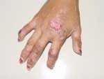
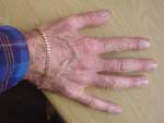
Porphyric skin disease has a characteristic appearance. Lesions are found only in sun-exposed areas: typically the backs of the hands, the forearms and to a lesser extent, the back of the neck and the feet in people who wear open shoes or sandals. In the acute setting, lesions consist of fluid-filled blisters and small erosions (Click on the images). Typically these are few in number, with only 3 or 4 small fresh lesions at any time. A common associated finding are milia: pinpoint deposits of a whitish, waxy material within the skin. Chronic appearances in most cases consist of small marks, the residue of the acute lesions, which tend to be either lighter or darker than surrounding skin. These may persist for months or years, gradually fading as time passes. In most cases of variegate porphyria and porphyria cutanea tarda, skin disease is fairly subtle.
Photomutilation
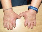
Severe skin disease is a feature of untreated porphyria cutanea tarda and of the exceedingly rare disorders congenital erythropoietic porphyria (See Unusual forms of porphyria) and the homozygous forms of variegate porphyria and porphyria cutanea tarda (also known as hepatoerythropoietic porphyria). In these disorders, the skin in sun-exposed areas becomes increasingly disfigured with pigmented blotches, coarsening and premature aging of the skin, hypertrichosis and eventually photomutilation with sclerodactyly and contraction and ultimately loss of appendages such as eyelids, nose, ears and lips.
The image on the left shows the hands of a ten-year old child with homozygous variegate porphyria: note the chronic skin damage and mutilating changes in the fingers.
Pathogenesis
The phenomenon of fluorescence
Porphyrin molecules exhibit the property of fluorescence. Light is absorbed at ultraviolet wavelengths centred around 406 nm. The trapped energy promotes electrons to a higher-energy state. As the electrons subsequently relax to the low-energy state, they re-radiate the additional energy at red wavelengths-typically about 630 nm. It is believed that these energy fluxes, occurring within the skin, underlie the photodermatitis characteristic of the porphyrias. There are thus two factors necessary for the pathogenesis of the skin lesions: excess porphyrins within the skin, and exposure to light. Frequently a third factor—trauma—is present: it is often noted that the skin breaks easily after minor trauma such as knocks and scrapes, and then heals unusually slowly.
Ultraviolet light and photosensitivity
Not all wavelengths of light have the same damaging effect on the skin. It is principally the long-wavelength ultraviolet light (UVA), of wavelength approaching 400 nm, that is most damaging. Note that these wavelengths are considerably longer than those which cause sunburn—UVB. Even longer wavelengths, in the visible spectrum, can be damaging in porphyria. This has important consequences for the prevention of porphyrin skin disease: conventional sunblocks, which screen out UVB rather than UVA, are unhelpful in preventing porphyric skin damage.
Porphyria Cutanea Tarda
The skin disease of porphyria cutanea tarda (Read Porphyria cutanea tarda) is similar to that of variegate porphyria, and in milder cases the two cannot be distinguished clinically. In some cases of porphyria cutanea tarda the skin disease is more severe, with large, ugly blisters and open sores on the hands and face , darkening and coarsening of the skin, hypertrichosis and sclerodactyly. Thickening and tightening of the skin of the face and hands may be so marked as to suggest a diagnosis of scleroderma, but unlike scleroderma, the damage is confined to sun-exposed areas.
Erythropoietic Protoporphyria
The skin disease of erythropoietic protoporphyria takes a different form: an unusual syndrome of immediate, severe photosensitivity, felt by the patient as warmth, burning, stinging and pain in sun-exposed areas (Read Erythropoietic protoporphyria). Typically patients have a threshold: and beyond a set period of exposure (usually 30 minutes), will learn to expect the symptoms to begin. When seen immediately after sun exposure, the skin may be erythematous and oedematous. Unlike the other porphyrias, the patients do not develop erosions and blisters, but rather an unusual but characteristic pattern of thickening and coarsening of the skin over the bridge of the nose and the knuckles.
Relationship Between Light Exposure and the Acute Attack
There is no such relationship. Some patients mistakenly believe that exposure to sunlight will precipitate critical complications such as the acute attack and paralysis. This is totally erroneous. The worst consequence of sun-exposure is aggravated skin disease.
Management of Skin Disease
Read the following: Management of skin disease.


