PCR
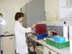
DNA is extracted from blood. By a process called the polymerase chain reaction (PCR), a short section covering the appropriate area of the gene suspected of carrying a mutation is greatly amplified—multiplied up in large quantities. This is performed automatically by a PCR cycler, a machine as shown.
Restriction Digest ASSAY
The amplified DNA (known as the PCR product) is then applied to a special plate known as a gel. Some of the sample is treated with a special
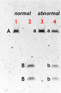
enzyme known as a restriction enzyme which cuts the PCR product at a specific point into two shorter pieces. This point is carefully chosen so that, if the mutation under investigation is present, cutting does not take place. The single piece corresponding to the product is therefore replaced by two smaller pieces. An electric current is applied for 20 minutes which causes the DNA fragments to move down through the gel at a speed corresponding to their size. A stain is applied which allows us to see the pieces. If the product was not cut, then a single fragment is seen, telling us that the mutation was present. Two lines lower down corresponding to the two cut pieces which, being smaller, travel further, tell us that the mutation was absent - the patient was normal. (Click on the image for an annotated picture.)
We can even confirm that the patient is
heterozygous - such a patient has one normal allele and one mutated allele (from each of two parents respectively). In this event, one sees three lines on the gel—two lines corresponding to the cut product, and one corresponding to the uncut product.
The image on the left is of a restriction digest test for the common R59W mutation in South African patients with VP. Where three lines are present, the test is positive forVP. Where two lines are present, the test is negative.
DNA Sequencing
The restriction digest assay is a quick and simple test which works well for mutations already known and characterised, such as the R59W mutation for
VP in South Africa. However, where the presence of porphyria is proven using
biochemical tests but we cannot show the presence of one of the known mutations, then one has to set about identifying this new mutation. In this case, the PCR product is subjected to automatic sequencing. This produces a graph which identifies the sequence of
bases, and allows us to recognise mutations. The example on the right shows two overlapping peaks in the middle of the graph. These represent two different bases occupying the same spot: one (inherited from one parent) is normal, the other (inherited from the other parent) is a mutation.


