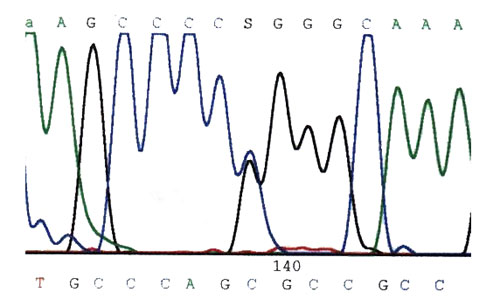
DNA has been extracted from blood, and part of the gene for the area of interest amplified by the PCR reaction. The sequence of bases is plotted out by the automatic sequencer and are identified by the letters at the top : AGCCC(S)GGGCAA (representing alanine,cytosine and guanine). "S" signifies an unknown, and one can clearly see that there are two different bases occupying the same spot - hence the superimposed blue and black lines. This is the actual mutation. In fact these are a C (blue) and a G (black) base. One has been inherited from mother, the other from father, and in the case shown, this single base substitution is enough to result in an inactive, mutant enzyme with resultant porphyria in the patient. One cannot tell from the sequence which is the abnormal base - this is deduced by comparing the sequence with that taken from normal people, and unaffected family members.